AI harnessed for early identification of cardiovascular problems

Roula Khalaf, Editor of the FT, selects her favourite stories in this weekly newsletter.
Cardiovascular disease is the world’s biggest killer, responsible for almost 18m deaths a year, according to the World Health Organization. But what if your likelihood of suffering from heart disease could be identified a decade in advance, enabling you to take preventive measures?
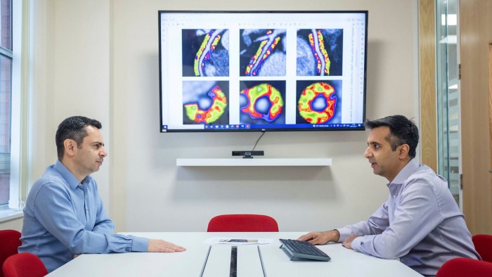
That is the promise of new medical imaging developed at the University of Oxford that received EU approval in 2021 and has begun to be used at several hospitals in the UK. The technology, CaRi-Heart, detects normally invisible signs of inflammation around coronary arteries from routine heart scans and uses artificial intelligence to identify the problems that could cause a heart attack.
Charalambos Antoniades, professor of cardiovascular medicine at Oxford, is the imaging expert behind the development, which received early backing from the British Heart Foundation (BHF) and has been spun off into a company, Caristo Diagnostics. “We have developed a method that takes the scans, uses AI to analyse not only the arteries of the heart but also the area around the arteries, and this tells you whether you will have a heart attack in the next eight to 10 years,” Antoniades says. This gives the patient time to adopt preventive measures such as taking statins or improving their diet.
Cheerag Shirodaria, Caristo co-founder and chief executive, says the technology — which is cheap, non-invasive and does not requires any special software — could be particularly beneficial for developing countries that are experiencing an “explosion” of heart disease. “What Covid has taught us is that we need to prevent disease,” he says. “We’ve spent far too much money on patients once they’ve already got an established disease when they’re in hospital. If we spent more on preventing disease . . . we would save a ton of money in the long run.”
But, even when backed by clinical evidence, it can be a challenge to put this kind of technology into practice, says Antoniades. “You’re talking about saving costs over the horizon of a decade,” he says. “In most countries, decisions are made by employees or politicians who have a short [decision-making] lifespan. That’s why prognostic and preventive medical devices are hard to get adopted.”
Another development, which Antoniades has written on in the European Heart Journal, is the potential to diagnose heart disease from facial photos, as outlined in a Chinese study. It was already known that features such as male pattern baldness and cholesterol deposits around the eyes are associated with increased heart risk, but AI makes it easier to analyse this data without human intervention. The prospect could be significant for health systems that cannot afford effective screening programmes. Antoniades says it raises concerns about data protection and rising insurance premiums for those identified as at risk from heart problems.
Another imaging advance that has benefited from BHF funding comes from Declan O’Regan, professor of imaging sciences at Imperial College London. He uses AI to build 3D models of the heart from MRI scans to better understand the causes of heart failure. “It’s about understanding the heart as a 3D piece of engineering, rather than relying on measures like ejection fraction — the percentage of blood that it pumps out each time. Those measures are crude, approximate and subjective,” he says. “They’re weak predictors of outcomes. By building a really immersive 3D model, we can get much more useful information.”
O’Regan and his colleagues have analysed thousands of scans from the UK Biobank database, helping them build “super-detailed digital maps” of the heart to identify which patients might benefit from earlier, more aggressive treatment. “It may mean also, conversely, being able to reassure some patients that they don’t need more aggressive therapy,” O’Regan says.
“People have their scan, the computers analyse their beating heart while they’re having that scan and, before they’ve even got out of the scanner, it will have analysed those images and provided the radiologist with a sliding scale of what degree of risk that patient is at and what sort of treatments might benefit them.”
Many of these AI technologies could change how care is given, says Julie Hart, director of strategic and industry partnerships at the Oxford Academic Health Science Network, which is helping Caristo secure NHS adoption of its technology. “It’s almost a Star Trek scenario,” she says, “being able to look at people with a scanner and use AI to give a risk stratification or diagnosis . . . we really are going into the next generation of healthcare delivery.”

Comments